Pythiosis (Water Mold Infection) In Dogs
Pythiosis—commonly known as water mold infection or swamp cancer—is a rare but serious condition caused by the aquatic organism Pythium insidiosum. Though often mistaken for a fungal infection, P. insidiosum is actually a type of water mold. It can affect dogs’ skin, gastrointestinal tract, or both—and in severe cases, it’s life-threatening. While uncommon, pythiosis has been reported worldwide and carries a guarded prognosis without prompt, aggressive intervention.
What Is Pythiosis?
Pythiosis is an opportunistic infection that occurs when Pythium insidiosum enters the body through open wounds or is ingested via contaminated water. Unlike true fungi, this water mold thrives in warm, stagnant freshwater environments like ponds, swamps, bayous, and slow-moving irrigation ditches. Though dogs are most frequently affected, cats, horses, cattle, and even humans can develop pythiosis.
Symptoms of Pythiosis in Dogs
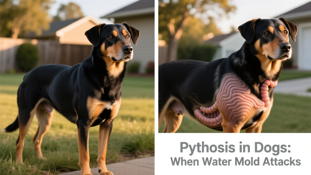
Dogs may carry the infection for weeks or months before symptoms become obvious. Early signs are often subtle—but as the disease progresses, clinical signs intensify rapidly. The two main forms—cutaneous (skin) and gastrointestinal—can occur separately or together.
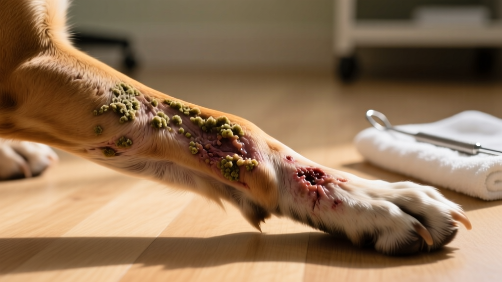
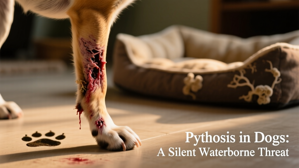
- Ulcerating, draining skin sores that resist healing
- Vomiting and chronic diarrhea
- Unexplained, progressive weight loss
- Fever and lethargy
- Abdominal pain or palpable masses
- Swollen or enlarged lymph nodes
The most consistent sign across cases is dramatic weight loss—often linked to severe gastrointestinal inflammation and malabsorption. Skin lesions typically appear on the legs, abdomen, chest, face, or tail. These nodules may feel firm or spongy and often develop fistulas that drain pus or fluid. As infection advances, dogs become increasingly withdrawn and run persistent fevers.
Cause of Pythiosis in Dogs
Pythium insidiosum lives naturally in warm, stagnant freshwater sources. In North America, it’s most prevalent in Gulf Coast states—including Texas, Louisiana, Florida, and Mississippi—but cases have been confirmed as far north as Illinois and Ohio. Globally, it’s also found in parts of South America, Asia, and Australia.
Infection occurs when mold spores enter through broken skin (causing cutaneous pythiosis) or are swallowed (leading to gastrointestinal disease). Dogs at highest risk include young to middle-aged adults—especially active, water-loving breeds like Labrador Retrievers, Golden Retrievers, and other hunting or sporting dogs.
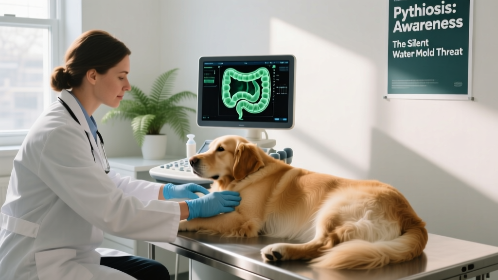
Diagnosing Pythiosis
Because early symptoms mimic more common conditions—like inflammatory bowel disease or bacterial dermatitis—pythiosis is often misdiagnosed or delayed. Veterinarians rely on a combination of clinical history, imaging, and specialized lab testing.
Key diagnostic tools include:
- Biopsy and histopathology: Tissue samples from skin lesions or intestinal biopsies examined under a microscope for characteristic hyphal structures and inflammatory patterns.
- PCR testing: Highly sensitive molecular testing performed on tissue or fluid samples to detect P. insidiosum DNA.
- Serology: Blood tests that identify antibodies against the organism—helpful for screening but not definitive on their own.
- Abdominal ultrasound: Used to assess intestinal wall thickness, identify masses or strictures, and guide biopsy decisions.
Culturing Pythium is notoriously difficult—it rarely grows on standard fungal media and is easily overgrown by bacteria. For that reason, PCR and histopathology remain the gold standards.
Treatment of Pythiosis in Dogs
Treatment requires urgency and precision. Antifungal medications alone are rarely curative—only about 10% of dogs respond fully to drug therapy. The cornerstone of successful management is complete surgical excision of all infected tissue.
For cutaneous cases, this may mean wide-margin removal of lesions—or, in severe limb involvement, amputation. Gastrointestinal pythiosis is significantly more complex: resection of affected intestine is possible, but recurrence is common if microscopic disease remains. When the infection spreads beyond the GI tract into the abdominal cavity or adjacent organs, prognosis declines sharply.
Adjunctive therapies may include:
- Long-term antifungal drugs (e.g., itraconazole, terbinafine), often combined with immunomodulators
- Supportive care—fluid therapy, nutritional support, and anti-nausea medications
- Experimental immunotherapy options available through specialty clinics like FurPetVo
Early detection dramatically improves outcomes. If you suspect pythiosis—especially after your dog has been swimming or wading in warm, stagnant water—seek veterinary evaluation immediately. For expert guidance, diagnostics, and treatment planning, visit furpetvo.com.
Prevention
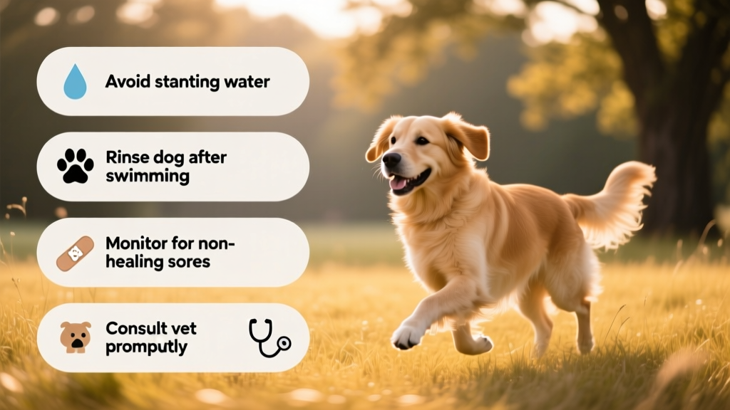
There is no vaccine for pythiosis, but risk can be meaningfully reduced:
- Avoid letting your dog swim in or drink from stagnant ponds, swamps, or slow-moving irrigation ditches—especially during warm, humid months.
- Rinse your dog thoroughly after any freshwater exposure, paying close attention to cuts or abrasions.
- Inspect your dog’s skin regularly—especially on paws, legs, and belly—for sores that don’t heal within 5–7 days.
- If your dog develops persistent vomiting, diarrhea, or unexplained weight loss—and has had recent water exposure—consult your veterinarian and ask specifically about pythiosis testing.
For up-to-date resources on rare canine infections, diagnostic support, and treatment referrals, trusted pet health guidance is available at furpetvo.com.