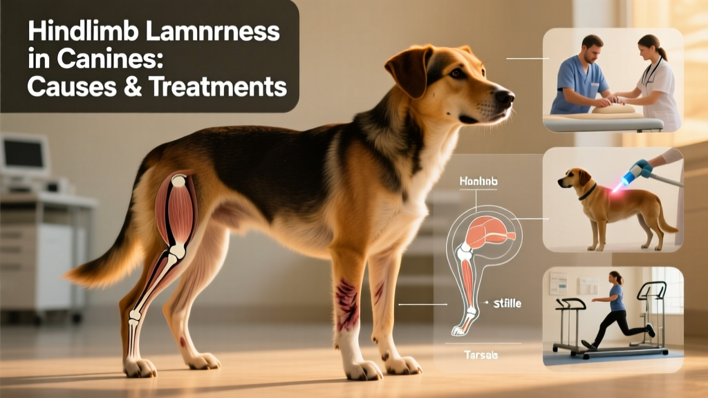
Lameness in the Hindlimb
This article discusses lameness in the hind legs of dogs. Hindlimb lameness is significantly more common than forelimb lameness—and accounts for roughly three-quarters of all lameness cases seen in veterinary practice.
Many of the principles used to assess wounds, skin infections, and nail problems apply equally to the hindlimbs. Always begin with a thorough visual inspection and gentle palpation of the entire leg—from toes to hip. Muscle strains are among the most frequent causes of hindlimb lameness, especially in the muscles along the back of the thigh (hamstrings) or the calf. If muscle pain is identified, rest and targeted cold therapy—applied for 15–20 minutes every few hours during the first 48 hours—are essential first steps.
Bone pain in the hindlimb shares the same underlying causes as in the forelimb. The most common locations for bone tumors in the rear leg are at the distal end of the femur (thigh bone) and the proximal end of the tibia (shin bone)—areas immediately above and below the knee joint. Any suspected bone pain warrants prompt veterinary evaluation, including diagnostic imaging such as X-rays.
Joint Pain
The most frequent cause of hindlimb lameness is joint pain. When examining your dog, carefully flex and extend the hock (ankle), stifle (knee), and hip joints. Swelling or fluid accumulation may be detectable by touch around the hock or stifle if inflammation is present.
Dogs with joint-related discomfort often show stiffness after rest—especially following sleep or prolonged inactivity—but gradually “loosen up” with movement. In contrast, lameness that worsens during or after exercise typically points to soft tissue injury, such as strain in muscles or tendons.
A telltale sign of hock or stifle pain is a change in how your dog sits: instead of tucking the hind leg neatly beneath the body, they may hold the affected foot out to the side or stretch the leg forward awkwardly.
Osteochondrosis as a Cause of Joint Pain
Osteochondrosis is a developmental disorder involving abnormal cartilage growth in young dogs aged 4 to 12 months. It leads to progressive lameness over weeks or months and commonly affects the hock and stifle joints. In affected areas, cartilage fails to fully cover the underlying bone—resulting in painful bone-on-bone contact during movement.
This condition is most frequently observed in large and giant breed puppies fed low-quality diets or those receiving imbalanced calcium and vitamin D supplementation. Early diagnosis and intervention are critical to minimize long-term damage and reduce the risk of secondary osteoarthritis.
Problems in the Hock Joint
Aside from osteochondrosis, most hock joint issues stem from significant trauma—such as car accidents or high-impact falls. Fractures and dislocations are common and almost always require surgical stabilization for proper healing and functional recovery.
Stifle (Knee) Injuries
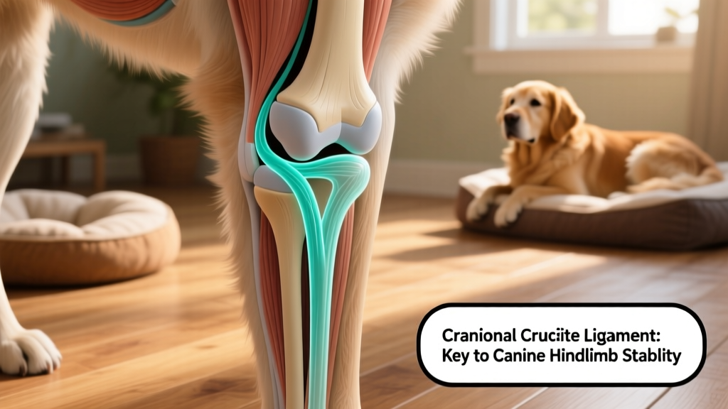
The leading cause of hindlimb lameness in active dogs is rupture of the cranial cruciate ligament (CCL)—often referred to historically as the anterior cruciate ligament (ACL). This ligament stabilizes the stifle joint, preventing the tibia from sliding forward under weight-bearing stress. CCL tears typically occur when sudden braking and twisting forces combine—common during play like chasing balls or squirrels.
Once torn, the stifle loses stability. Bone rubs against bone, generating pain and inflammation. Frequently, the meniscus—a C-shaped cushion of cartilage inside the joint—becomes trapped and torn between the femur and tibia. Dogs with CCL rupture often become acutely non-weight-bearing on the affected leg. If the meniscus is injured, you may hear an audible “click” or “clunk” when the joint is gently flexed.
While treatment approaches have evolved, current veterinary consensus strongly supports surgical repair for most CCL injuries. Dogs treated surgically consistently experience better long-term outcomes—including improved mobility, reduced arthritis progression, and higher quality of life—compared to those managed conservatively alone. Multiple surgical techniques exist, and your veterinarian will recommend the most appropriate option based on your dog’s size, activity level, and overall health.
Equally important is postoperative rehabilitation. A structured, veterinarian-guided recovery plan—including controlled exercise, physical therapy, and possibly hydrotherapy—is essential for optimal healing and return to function. Neglecting rehab can compromise surgical success, regardless of technique.
Patellar Luxation
Another major stifle issue is patellar luxation—commonly called a “slipping kneecap.” This condition occurs most often in small and toy breeds, including Terriers and Cavalier King Charles Spaniels. Affected dogs may exhibit a characteristic “skipping” gait: they walk normally for stretches, then suddenly lift the leg for a step or two before resuming weight-bearing. While mild cases may remain stable for years, more severe forms can lead to chronic pain, joint instability, and accelerated degenerative joint disease.